
Sidoinnehåll
Inledning
Idag är det allt vanligare att patienter som kommer till PCI-labbet redan har genomgått en diagnostisk kranskärls-CT. Detta ger oss ett värdefullt verktyg som bör utnyttjas för att optimera planeringen av PCI. CTCA:s utveckling har lett till AI-baserade system för integration i angiolabbets PCI-arbetsflöde.1
En enkät bland PCI-operatörer i P4-studien (CT-guidad vs. IVUS-guidad PCI) visade att pre-procedural planering värderas mycket högt, särskilt bedömning av placksammansättning och kalciumkarakteristik, kalciumbåge ansågs vara den mest användbara informationen.
Undersökningen lyfte även fram betydelsen av att bedöma area at risk vid bifurkations-PCI och förväntad kalciumtäthet som en framtida nyckelfaktor vid CT-guidad PCI.2
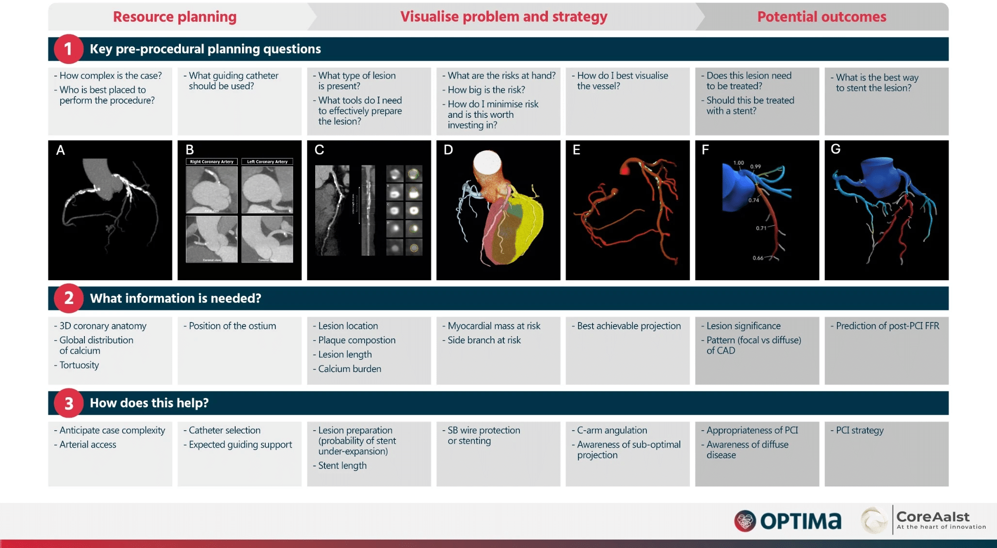
Sammanfattning av CCTA-guidad PCI:2
Val av guidekateter
Koronar-CT kan i axiella vyer visa koronarkärlens avgångsvinklar och därmed hjälpa till att välja optimal guidande kateter inför PCI. Till exempel kan en anterior avgång av höger koronarartär göra att klassiska katetrar som Judkins right ger otillräckligt stöd, och istället tala för att använda en Amplatz left-kateter.
Genom att planera kateterval i förväg kan koronar-CT bidra till att minska kontrastmängd, procedurtid och behov av kateterbyten. CT är också förstahandsmetod för att identifiera kärlavgångar vid kranskärlsanomali och bypassgraft.1
Planering för CABG-opererade patienter
Koronar-CT är en värdefull metod för patienter med tidigare bypassoperation (CABG) som planeras för koronarangiografi.
Den ger viktig information som kan förbättra proceduren och minska risker.1
Koronar-CT kan visa:
- Antalet bypassgraft.
- Positionen för ostiet och målkärl.
- Patency och eventuella lesioner i graften.3
Fördelar:
- Bättre materialval och accessplanering.
- Kortare procedurtid och färre komplikationer.
- Ökad användning av radial access och högre patientnöjdhet (BYPASS-CTCA-studien).4
Myocardial Mass at Risk
Myokardmassa som försörjs av varje kärl kan automatiskt beräknas från koronar-CT med Voronoi–5 6 eller CFD-baserade7 algoritmer, något som är särskilt användbart vid PCI-planering av bifurkationer.1
Analysen skiljer inte mellan viabelt och icke-viabelt myokard, vilket dock kan lösas med kombinerad CT-perfusion.1
- Voronoi: 5 6
- Enkel geometrisk indelning baserat på närmsta kärl.
- Beräknar försörjd myokardmassa, men inte blodflöde.
- CFD (Computational Fluid Dynamics): 7
- Simulerar blodflöde och tryck i kärlen.
- Använder myokardmassan för att beräkna flöde och trans-lesionella tryckgradienter.
- Kopplar försörjd myokardmassa till FFR och ischemi.
- Ger mer exakt fysiologisk information men är mer resurskrävande.
PCI för förkalkade lesioner
CT:s höga känslighet för kalcium erbjuder en unik möjlighet att bedöma och kartlägga förkalkningars omfattning och svårighetsgrad.8 9
CTCA kan bidra med:
- Kartläggning av kalciumbörda, utbredning, tjocklek och längd, även i bifurkationer
- Identifiering av koncentriska kalciumringar.
- Vägledning för val av plackmodifieringsteknik (cutting balloon, IVL, RA, OA).
- Tidig förutsägelse av behovet av rotationsaterektomi baserat på per-lesion-kalciumscore och kalciumbåge.10 11 12
Begränsningar:
- Kan ännu inte skilja mellan ytlig och djup kalk.1
- CTCA kan i dagsläget inte särskilja nodulära från andra typer av förkalkningar.1
Se även sidan PCI-behandling av förkalkade kranskärl
Relaterade sidor
Litteratur
Computed tomographic angiography in coronary artery disease (2023)13
SCCT 2021 Expert Consensus Document on Coronary Computed Tomographic Angiography: A Report of the Society of Cardiovascular Computed Tomography 14
Pre-procedural planning of coronary revascularization by cardiac computed tomography: An expert consensus document of the Society of Cardiovascular Computed Tomography (2022)15
Coronary CT Angiography in the Cath Lab: Leveraging Artificial Intelligence to Plan and Guide Percutaneous Coronary Intervention1
Clinical utility of coronary CT angiography to guide PCI: a survey among P4 investigators2
Implementing Coronary Computed Tomography Angiography in the Catheterization Laboratory 16
Kalk
Percutaneous coronary intervention for calcified and resistant lesions 8
Management strategies for heavily calcified coronary stenoses: an EAPCI clinical consensus statement in collaboration with the EURO4C-PCR group9
Quantification of calcium burden by coronary CT angiography compared to optical coherence tomography 11
CABG
Computed Tomography Cardiac Angiography Before Invasive Coronary Angiography in Patients With Previous Bypass Surgery: The BYPASS-CTCA Trial4
Last Updated on May 3, 2025 by Christian Dworeck
- ICD-söktjänst: - August 23, 2025
- Ny sida: Var blir jag citerad? - August 13, 2025
- Ny sida: PCI vid förkalkade kranskärl – teknik, tips och evidens - May 6, 2025
- Ohashi H, Bouisset F, Buytaert D, Seki R, Sonck J, Sakai K, Belmonte M, Kitslaar P, Updegrove A, Amano T, Andreini D, De Bruyne B, Collet C. Coronary CT Angiography in the Cath Lab: Leveraging Artificial Intelligence to Plan and Guide Percutaneous Coronary Intervention. Interv Cardiol. 2023 Nov 23;18:e26. doi: 10.15420/icr.2023.12. PMID: 38125928; PMCID: PMC10731535. [↩] [↩] [↩] [↩] [↩] [↩] [↩] [↩]
- Stalikas N, Bouisset F, Mizukami T, Tajima A, Munhoz D, Ikeda K, Sonck J, Wyffels E, Wilgenhof A, Astudillo P, Trabattoni D, Montorsi P, Zivelonghi C, Agostoni P, Scott B, Vermeersch P, Gallinoro E, Monizzi G, Andreini D, Vandeloo B, Lochy S, Argacha JF, Støttrup NB, Maeng M, Engstrøm T, Arslani K, Olsen NT, Ando H, Amano T, Ohashi H, Jeremias A, Ali Z, Shlofmitz E, Sakai K, Spratt JC, Brilakis ES, Sandoval Y, Stefanini G, Bagnall A, Purcell I, Edes IF, De Bruyne B, Collet C. Clinical utility of coronary CT angiography to guide PCI: a survey among P4 investigators. Int J Cardiovasc Imaging. 2025 Feb 19. doi: 10.1007/s10554-025-03323-y. Epub ahead of print. PMID: 39971841. [↩] [↩] [↩] [↩]
- Barbero U, Iannaccone M, d’Ascenzo F, Barbero C, Mohamed A, Annone U, Benedetto S, Celentani D, Gagliardi M, Moretti C, Gaita F. 64 slice-coronary computed tomography sensitivity and specificity in the evaluation of coronary artery bypass graft stenosis: A meta-analysis. Int J Cardiol. 2016 Aug 1;216:52-7. doi: 10.1016/j.ijcard.2016.04.156. Epub 2016 Apr 22. PMID: 27140337. [↩]
- Jones DA, Beirne AM, Kelham M, Rathod KS, Andiapen M, Wynne L, Godec T, Forooghi N, Ramaseshan R, Moon JC, Davies C, Bourantas CV, Baumbach A, Manisty C, Wragg A, Ahluwalia A, Pugliese F, Mathur A; BYPASS-CTCA Trial Committees and Investigators. Computed Tomography Cardiac Angiography Before Invasive Coronary Angiography in Patients With Previous Bypass Surgery: The BYPASS-CTCA Trial. Circulation. 2023 Oct 31;148(18):1371-1380. doi: 10.1161/CIRCULATIONAHA.123.064465. Epub 2023 Sep 29. PMID: 37772419; PMCID: PMC11139242. [↩] [↩]
- Ide S, Sumitsuji S, Yamaguchi O, Sakata Y. Cardiac computed tomography-derived myocardial mass at risk using the Voronoi-based segmentation algorithm: A histological validation study. J Cardiovasc Comput Tomogr. 2017 May-Jun;11(3):179-182. doi: 10.1016/j.jcct.2017.04.007. Epub 2017 Apr 18. PMID: 28431861. [↩] [↩]
- van Driest FY, Bijns CM, van der Geest RJ, Broersen A, Dijkstra J, Jukema JW, Scholte AJHA. Correlation between quantification of myocardial area at risk and ischemic burden at cardiac computed tomography. Eur J Radiol Open. 2022 Mar 31;9:100417. doi: 10.1016/j.ejro.2022.100417. PMID: 35402660; PMCID: PMC8983940. [↩] [↩]
- Taylor CA, Fonte TA, Min JK. Computational fluid dynamics applied to cardiac computed tomography for noninvasive quantification of fractional flow reserve: scientific basis. J Am Coll Cardiol. 2013 Jun 4;61(22):2233-41. doi: 10.1016/j.jacc.2012.11.083. Epub 2013 Apr 3. PMID: 23562923. [↩] [↩]
- Pesarini G, Hellig F, Seth A, Shlofmitz RA, Ribichini FL. Percutaneous coronary intervention for calcified and resistant lesions. EuroIntervention. 2025 Apr 7;21(7):e339-e355. doi: 10.4244/EIJ-D-24-00195. PMID: 40191879; PMCID: PMC11956026. [↩] [↩]
- Barbato E, Gallinoro E, Abdel-Wahab M, Andreini D, Carrié D, Di Mario C, Dudek D, Escaned J, Fajadet J, Guagliumi G, Hill J, McEntegart M, Mashayekhi K, Mezilis N, Onuma Y, Reczuch K, Shlofmitz R, Stefanini G, Tarantini G, Toth GG, Vaquerizo B, Wijns W, Ribichini FL. Management strategies for heavily calcified coronary stenoses: an EAPCI clinical consensus statement in collaboration with the EURO4C-PCR group. Eur Heart J. 2023 Nov 1;44(41):4340-4356. doi: 10.1093/eurheartj/ehad342. PMID: 37208199. [↩] [↩]
- Sekimoto T, Akutsu Y, Hamazaki Y, Sakai K, Kosaki R, Yokota H, Tsujita H, Tsukamoto S, Kaneko K, Sakurai M, Kodama Y, Li HL, Sambe T, Oguchi K, Uchida N, Kobayashi S, Aoki A, Gokan T, Kobayashi Y. Regional calcified plaque score evaluated by multidetector computed tomography for predicting the addition of rotational atherectomy during percutaneous coronary intervention. J Cardiovasc Comput Tomogr. 2016 May-Jun;10(3):221-8. doi: 10.1016/j.jcct.2016.01.004. Epub 2016 Jan 13. PMID: 26811266. [↩]
- Monizzi G, Sonck J, Nagumo S, Buytaert D, Van Hoe L, Grancini L, Bartorelli AL, Vanhoenacker P, Simons P, Bladt O, Wyffels E, De Bruyne B, Andreini D, Collet C. Quantification of calcium burden by coronary CT angiography compared to optical coherence tomography. Int J Cardiovasc Imaging. 2020 Dec;36(12):2393-2402. doi: 10.1007/s10554-020-01839-z. Epub 2020 Nov 17. PMID: 33205340. [↩] [↩]
- Kurogi K, Ishii M, Nagatomo T, Tokai T, Kaichi R, Takae M, Mori T, Komaki S, Yamamoto N, Tsujita K. Mean density of computed tomography for predicting rotational atherectomy during percutaneous coronary intervention. J Cardiovasc Comput Tomogr. 2023 Mar-Apr;17(2):120-129. doi: 10.1016/j.jcct.2023.02.002. Epub 2023 Feb 10. PMID: 36775780. [↩]
- Serruys PW, Kotoku N, Nørgaard BL, Garg S, Nieman K, Dweck MR, Bax JJ, Knuuti J, Narula J, Perera D, Taylor CA, Leipsic JA, Nicol ED, Piazza N, Schultz CJ, Kitagawa K, Bruyne B, Collet C, Tanaka K, Mushtaq S, Belmonte M, Dudek D, Zlahoda-Huzior A, Tu S, Wijns W, Sharif F, Budoff MJ, Mey J, Andreini D, Onuma Y. Computed tomographic angiography in coronary artery disease. EuroIntervention. 2023 Apr 3;18(16):e1307-e1327. doi: 10.4244/EIJ-D-22-00776. PMID: 37025086; PMCID: PMC10071125. [↩]
- Narula J, Chandrashekhar Y, Ahmadi A, Abbara S, Berman DS, Blankstein R, Leipsic J, Newby D, Nicol ED, Nieman K, Shaw L, Villines TC, Williams M, Hecht HS. SCCT 2021 Expert Consensus Document on Coronary Computed Tomographic Angiography: A Report of the Society of Cardiovascular Computed Tomography. J Cardiovasc Comput Tomogr. 2021 May-Jun;15(3):192-217. doi: 10.1016/j.jcct.2020.11.001. Epub 2020 Nov 20. PMID: 33303384; PMCID: PMC8713482. [↩]
- Andreini D, Collet C, Leipsic J, Nieman K, Bittencurt M, De Mey J, Buls N, Onuma Y, Mushtaq S, Conte E, Bartorelli AL, Stefanini G, Sonck J, Knaapen P, Ghoshhajra B, Serruys P. Pre-procedural planning of coronary revascularization by cardiac computed tomography: An expert consensus document of the Society of Cardiovascular Computed Tomography. J Cardiovasc Comput Tomogr. 2022 Nov-Dec;16(6):558-572. doi: 10.1016/j.jcct.2022.08.003. Epub 2022 Aug 22. PMID: 36008263. [↩]
- Collet C, Sonck J, Leipsic J, Monizzi G, Buytaert D, Kitslaar P, Andreini D, De Bruyne B. Implementing Coronary Computed Tomography Angiography in the Catheterization Laboratory. JACC Cardiovasc Imaging. 2021 Sep;14(9):1846-1855. doi: 10.1016/j.jcmg.2020.07.048. Epub 2020 Nov 25. PMID: 33248968. [↩]